Finding a painless lump deep within a muscle or limb feels a lot like spotting an unusual anomaly in production logs. Because the swelling does not hurt, the easiest reaction is to brush it off as a pulled muscle, sports haematoma, or benign fatty cyst.
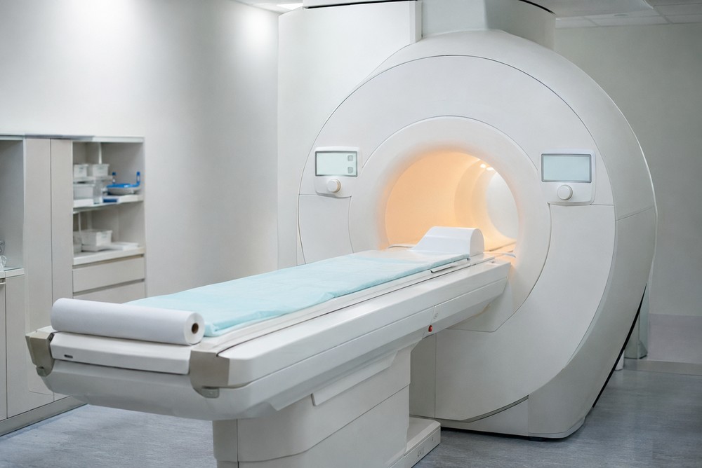
However, diagnosing deep soft tissue growths requires specialised imaging and histological evaluation rather than watchful waiting. If you are researching diagnostic pathways for suspected sarcoma in Singapore oncology specialists provide, reviewing how multidisciplinary sarcoma teams coordinate core biopsies, MRI staging, and limb-sparing resections gives you an objective foundation for your care. Establishing an accurate diagnosis early protects functional mobility while preventing incomplete initial surgeries.
The Biology of Deep Mesenchymal Growths
Connective tissue tumours arise from mesenchymal cells that develop into muscle, fat, nerves, blood vessels, and deep fibrous tissue. Unlike carcinomas that develop on surface linings, sarcomas form deep within compartments beneath muscular fascia.
Because muscular compartments stretch to accommodate expanding tissue, deep soft tissue masses often grow for months without causing pain. The tumour pushes outward, compressing normal cells into a pseudocapsule. While this boundary looks neat on physical inspection, microscopic projections of cancer cells routinely extend beyond the visible edge into adjacent muscle bundles.
Upfront Excision versus Core Needle Staging
Standard patient intuition almost always suggests that if you find an abnormal lump, you should cut it out immediately. The non-obvious trade-off is that attempting a quick shell-out excision without an upfront biopsy frequently contaminates healthy tissue compartments.
When a surgeon shells out an unbiopsied mass assumed to be a harmless lipoma, the cut breaches the tumour pseudocapsule, spilling cells into adjacent fascial planes. Choosing an image-guided core needle biopsy trades two weeks of diagnostic patience for surgical safety. Establishing the precise histological subtype beforehand allows a surgeon to plan an en-bloc resection with clear margins, sparing motor nerves and preventing radical secondary surgery.
The Benign Lipoma Assumption Mistake
The most common and expensive mistake patients make is assuming any painless, fleshy lump is a harmless lipoma or sports haematoma. People fall into this trap because benign fatty lumps are common, while soft tissue sarcomas represent less than one per cent of adult solid tumours.
In reality, simple fatty lipomas rarely develop deep beneath the muscular fascia, nor do they steadily expand over several months. Undergoing an unplanned excision at a non-specialist clinic without prior cross-sectional imaging transforms a localised mass into a contaminated surgical bed. Patients then require extensive revision surgery to resect the entire previous scar and wide surrounding margins, often sacrificing functional muscle groups to achieve clean borders.
Calculating Real Care Pathway Economics
Let us examine a concrete worked example comparing an unplanned local excision against an upfront multidisciplinary surgical pathway over twenty-four months. Consider an individual who undergoes a quick clinic excision for a six-centimetre thigh lump assumed to be a cyst, spending 4,500 dollars.
When histology reveals high-grade liposarcoma with positive margins, the patient requires emergency transfer to a specialist oncology centre. Repairing the contaminated compartment requires wide re-resection, reconstruction, and radiotherapy costing 38,000 dollars, plus physical therapy costing 4,000 dollars, bringing total spending to 46,500 dollars. In contrast, an initial pathway with contrast MRI, core biopsy, and planned single-stage resection costs 31,000 dollars total. Sourcing a specialised pathway upfront saves 15,500 dollars while preserving normal walking mechanics.
Core Habits for Managing Complex Staging
Navigating an uncertain soft tissue diagnosis requires establishing simple operational habits before consenting to any procedure. Treating your diagnostic workup like a structured audit keeps your care plan grounded in clear imaging.
Keep these practical operational habits in mind:
- Obtain dedicated contrast MRI before cutting: Insist on cross-sectional MRI imaging to evaluate depth, fascial boundaries, and proximity to major blood vessels.
- Align biopsy tracts with future incisions: Ensure your radiologist places the biopsy needle along the exact surgical line so the entire needle tract can be removed during definitive surgery.
- Request multidisciplinary tumour board review: Confirm that your case is reviewed jointly by musculoskeletal radiologists, pathologists, and surgical oncologists before finalising treatment.
A Clear Decision Rule for Deep Masses
You do not need an oncology qualification to determine when an unexplained lump requires specialised sarcoma evaluation. You can apply a straightforward decision rule called the five-centimetre fascia check during your mirror self-exam.
Examine the lump and check three distinct physical criteria: is the mass larger than five centimetres (roughly the size of a standard golf ball), does it sit deep beneath the muscle layer rather than moving freely in the skin, and has it grown noticeably over the past four weeks? If the lump meets even one of these criteria, mandate a formal MRI and an image-guided core biopsy before allowing anyone to cut or aspirate it.
Navigating a connective tissue diagnosis is ultimately about creating a calm strategy that prioritises anatomical precision over quick fixes. By avoiding unplanned shell-out surgeries, demanding cross-sectional imaging upfront, and following clear size rules, you protect both your functional mobility and your finances. Approach your care decisions with calm discipline, rely on verified surgical principles, and move forward with quiet confidence in your recovery plan.